How 3D Scanning Ex-Vivo Specimens Is Transforming Surgical Precision and Pathology Collaboration
Share
How 3D Scanning Ex-Vivo Specimens Is Transforming Surgical Precision and Pathology Collaboration
Imagine a world where surgeons don’t have to step out of the operating room to get critical pathological feedback, where tumor data is delivered instantly, accurately, and visually, helping guide life-saving decisions.
What once sounded like science fiction is rapidly becoming reality thanks to advanced 3D scanning technology.
Today’s operating rooms are increasingly embracing digital tools that bridge the gap between surgeons and pathologists.
Among these, ex-vivo 3D scanning of surgical specimens stands out as a breakthrough technique that enhances communication, reduces risks, and improves outcomes.
Surgeons and pathologists have traditionally relied on frozen section analysis and verbal back-and-forth during complex surgeries, a workflow that’s time-consuming, disruptive, and prone to error.
In the context of oncologic surgery, where every millimeter counts, delays or miscommunications can impact a patient’s prognosis.
But there’s a solution that’s gaining momentum in clinical settings: structured light 3D scanning of ex-vivo specimens, which allows teams to visualize anatomical surfaces and margins with remarkable detail and share that information instantly in the operating theater.
This article dives deep into how this technology works, why it’s accelerating surgical workflows, and what it means for patient care.
We’ll explore real case data, tangible benefits, challenges, and how tools like the EinScan Medixa scanner are helping medical teams collaborate with precision.
By the end, you’ll clearly understand why this blend of 3D scanning and specimen mapping could be a game-changer in modern medical practice and how you can bring this capability to your team.
The Traditional Challenge in Surgical Pathology
In many major surgical procedures, especially tumor resections, surgeons depend on intraoperative pathology to ensure complete removal of disease.
The standard workflow typically involves:
-
Resecting the specimen during surgery,
-
Sending it out for frozen section analysis (FSA) to a pathology lab,
-
Waiting for results before continuing or closing.
While effective, this workflow has notable limitations:
-
It interrupts the surgical flow.
-
Feedback is often delayed due to lab distances and communication lag.
-
Spatial context about where tissue was cut or sampled is lost.
-
Surgeons must interpret verbal descriptions and static photos to make decisions.
These factors contribute to longer OR time, increased costs, and the potential for ambiguity when interpreting pathology data.
Clear and immediate documentation of specimen anatomy has long been an unmet need in surgical pathology reporting.
That gap is exactly where 3D scanning steps in, such as capturing digital specimens and mapping margin data in real time.
What Is Ex-Vivo 3D Scanning and How Does It Work?
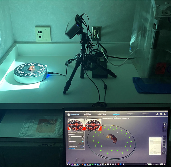
Ex-vivo 3D scanning means capturing the complete surface geometry of a freshly resected specimen outside the body using structured light scanners.
These devices project light patterns onto the specimen and calculate surface shape based on how those patterns deform, creating a high-resolution, three-dimensional digital model of the tissue.
A typical workflow might look like this:
-
After surgical resection, the fresh specimen is rinsed, patted dry, and placed on a turntable beneath a structured-light 3D scanner.
-
The scanner rotates and captures detailed surface topography in minutes (often ~8 minutes).
-
The resulting 3D data is exported to CAD or mesh-editing software.
-
Pathologists annotate the model to mark margin types, sampling sites, or tumor boundaries.
-
The annotated 3D specimen is displayed on screens in the OR and shared between teams in real time.
This process creates a digital twin of the specimen that reflects actual anatomy and allows interactive visualization, something no 2D photo or text report can match.
Why 3D Scanning Improves Surgical Collaboration
1. Real-Time Feedback in the Operating Room
Perhaps the biggest advantage is the ability to provide visual feedback without the surgeon leaving the OR.
By displaying a 3D model with annotations during frozen section analysis, both surgeon and pathologist stay in the loop together, interpreting margin status collectively. This drastically reduces wait times and miscommunication.
Instead of verbal descriptions or static photos, teams can point to exact anatomical landmarks, rotating and zooming in on digitally captured specimens. This clarity accelerates decision-making and enhances confidence during critical moments.
2. Accurate 3D Visualization of Surgical Margins
Knowing precisely where tumor tissue begins and ends isn’t just about completion, it’s about precision.
In standard workflows, tissue sections and margins are documented in writing or with flat photos. These references lack spatial depth and context.
A 3D specimen map, generated from a structured light scan, preserves the entire surface topology and marks every slice location accurately.
Surgeons have reported that features like frozen edges become clearly visible, reducing ambiguity in interpreting complex resection geometry and improving margin analysis clarity.
3. Improved Postoperative Documentation and Teaching
Digital specimen maps don’t just help during surgery, they serve as permanent records of a case’s morphology and surgical detail.
These 3D models can be archived and incorporated into teaching materials, quality reviews, and pathology reports where spatial understanding is critical.
They also support more accurate postoperative discussions and long-term case reviews, valuable for multidisciplinary teams, research, and training the next generation of clinicians.
4. Enhanced Communication With Visual Context
Unlike traditional 2D reporting, 3D models convey context visually and intuitively, making complex anatomy more understandable at a glance.
Whether it’s explaining results to a surgical team, trainees, or patients later on, detailed 3D scanning bridges communication gaps.
Studies and pilot implementations have shown that structured light 3D scanners improve documentation and help clinicians consistently interpret complex specimen geometry across disciplines.
Case Studies and Early Data
Clinical use cases and early research suggest that ex-vivo 3D scanning is not just plausible, it’s effective:
-
In a series of 40 cases, structured light 3D scanning captured detailed surface data in a median time of around 8 minutes per specimen, demonstrating real-world viability for intra-operative use.
-
Pilot studies highlight how 3D specimen maps improve documentation and can be integrated with conventional pathology reporting including custom annotations of tissue sampling and margin status.
-
Clinical reports note that virtual specimen maps can help surgeons and pathologists achieve better spatial context and understanding, which is difficult with traditional 2D documentation alone.
Are There Limitations?
Like any emerging technology, 3D scanning in pathology isn’t without challenges:
-
Training and Workflow Integration. Teams need training to operate scanners and annotate models efficiently.
-
Equipment and Space. Setting up a scanning station near pathology labs or ORs requires planning.
-
Data Management. Large 3D files need storage, processing software, and secure access.
However, scanned specimen data is relatively easy to integrate with existing digital pathology workflows, and costs are declining as adoption increases.
What the Future Holds
The integration of high-precision 3D scanning with surgical and pathology workflows is part of a broader trend toward digital transformation in medicine.
Advances in imaging technologies including augmented reality, intraoperative scanners, and digital pathology platforms are all pushing toward better, real-time clinical decision support.
As hospitals and surgical centers prioritize faster turnaround times and more integrated data flows, 3D specimen scanning will likely become a core part of advanced surgical pathology protocols.
Conclusion
Ex-vivo 3D scanning of surgical specimens is more than a technological novelty, it’s a practical solution to real clinical challenges.
By enabling detailed visualization, enhancing communication between surgeons and pathologists, and supporting intraoperative decision-making, structured light 3D scanning brings clarity and speed to a traditionally slow and fragmented workflow.
Whether you’re part of a surgical team, pathology department, or hospital administration looking to modernize clinical workflows, this technology is worth exploring.
Ready to Upgrade Your Surgical Workflow?
Explore the leading tools that make 3D scanning practical and powerful:

-
Try the EinScan Medixa — ideal for structured light scanning of limbs for orthotic and prosthetic devices.
-
Consider the EinScan Rigil — ideal for high resolution 3D scanning of surgical specimens and detailed 3D surface capture.
Frequently Asked Questions (FAQ)
1. How long does ex-vivo 3D scanning add to the surgical workflow?
In most clinical implementations, ex-vivo 3D scanning adds minimal time to the workflow. Structured light scanners can capture a complete specimen surface in approximately 5–10 minutes, often in parallel with routine frozen section preparation. Because scanning and annotation can occur while pathology analysis is underway, many teams report no net increase in operative time and in some cases, overall time savings due to faster, clearer communication.
2. How does 3D scanning improve margin assessment compared to traditional methods?
Traditional margin assessment relies on written descriptions, diagrams, or 2D photographs, which can lose spatial context. Ex-vivo 3D scanning preserves the full three-dimensional geometry of the specimen, allowing margins, slice locations, and sampling sites to be annotated directly on the digital model. This spatial accuracy helps surgeons understand exactly where a positive or close margin is located, reducing ambiguity and improving intraoperative decision-making.
3. Is ex-vivo 3D scanning compatible with existing pathology workflows?
Yes. Most structured light 3D scanning systems integrate smoothly with existing pathology processes. The scanned models can be exported to standard mesh or imaging software and linked to conventional pathology reports. While teams may need initial training and minor workflow adjustments, scanned specimen data is generally easy to incorporate into digital pathology, frozen section analysis, and postoperative documentation.
4. What types of surgeries benefit most from ex-vivo 3D specimen scanning?
Ex-vivo 3D scanning is particularly valuable in oncologic surgeries where margin status is critical, such as head and neck cancer resections, soft tissue tumors, breast surgery, and complex gastrointestinal or urologic cases. Any procedure where anatomical orientation, margin precision, or multidisciplinary communication is essential can benefit from enhanced 3D visualization.
5. Is patient data secure when using 3D scanned specimen models?
Patient data security is a key consideration. Most clinical 3D scanning workflows store specimen models on secure hospital servers or encrypted systems, similar to other medical imaging data. When properly implemented, 3D specimen files can comply with institutional data governance policies and healthcare privacy regulations, ensuring that digital models are used safely for clinical care, documentation, and education.